Warning signs of melanoma: A guide to clinical identification and skin health monitoring
Understanding Melanoma and the Importance of Early Detection
Melanoma represents the most serious form of skin cancer, originating in the pigment-producing cells known as melanocytes 5. While it accounts for less than 2% of all diagnosed skin cancers in the United States, it is responsible for the highest number of skin cancer-related deaths due to its tendency to metastasize to other parts of the body 5. Approximately 20% to 30% of these malignancies develop within existing moles, while the remaining 70% to 80% emerge on previously normal-looking skin 1. Early detection is a critical factor in improving patient outcomes, as the condition is often highly treatable when identified in its initial stages 1.
Because melanomas can manifest in diverse shapes, sizes, and colors, they may occasionally evade conventional warning signs, making routine self-examinations essential for high-risk individuals 1. Over 80% of melanomas are initially identified by patients, family members, or close acquaintances, underscoring the vital role of regular skin checks 2. Maintaining awareness of these changes is particularly important for individuals with a history of significant sun exposure, numerous moles, or a family history of skin cancer 2.
The ABCDE Rule: A Standard Clinical Framework
Dermatologists and healthcare providers utilize the ABCDE system as a standardized tool for identifying potential melanoma warning signs during physical examinations 2, 3. This mnemonic provides a structured approach for individuals to monitor their skin for concerning developments that require professional assessment 9.
| Letter | Clinical Sign | Description |
|---|---|---|
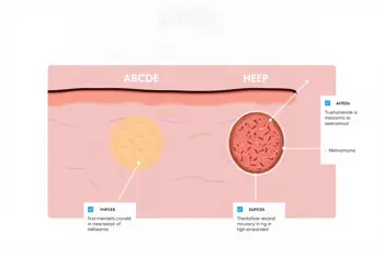
| A | Asymmetry | One half of the mole does not mirror the other half 2, 3. |
| B | Border | The edges are uneven, jagged, ragged, or blurred 2, 7. |
| C | Color | The lesion contains multiple shades, such as brown, black, tan, red, blue, or white 2, 3. |
| D | Diameter | The spot is typically larger than 6 mm, though smaller melanomas can exist 2, 6. |
| E | Evolving | The spot changes in size, shape, color, or behavior over weeks or months 2, 8. |
Each component of the ABCDE rule serves as a signal for potential malignancy, yet these features are not exhaustive. The presence of these characteristics does not confirm a diagnosis, but rather signals a need for formal medical evaluation 8. As clinical guidance emphasizes, any evolving mole-particularly one that is increasing in size or changing its appearance-should be treated as a priority for dermatological review 8.
Recognizing the Ugly Duckling Sign and Skin Variations
Beyond the formal ABCDE criteria, dermatologists emphasize the importance of the ugly duckling sign 1, 5. This concept refers to any spot that looks visibly different from the rest of a patient's moles 1, 2. Because most benign moles on an individual often share a similar appearance, a single lesion that stands out due to its unique color or architecture warrants professional attention, even if it does not strictly adhere to all components of the ABCDE rule 5.
Melanoma is not limited to typical presentations; while many are dark brown or black, others may manifest as red, pink, or even skin-colored growths that lack heavy pigmentation 6, 10. Furthermore, lesions may present with changes to their surface, such as the development of crusting, scaliness, bleeding, or the presence of a new lump or bump 1, 2. These textural changes are considered significant clinical indicators that require a professional biopsy to definitively rule out malignancy 5, 7.
Symptomatic Indicators Beyond Visual Changes
While visual changes often prompt a clinical visit, physical sensations associated with a skin lesion can also serve as warning signs. Symptoms such as persistent itching, tenderness, or unexplained pain in a specific mole should never be ignored, as these feelings can sometimes correlate with the growth of malignant cells 1, 2. Any sore that refuses to heal after a reasonable period is a recognized clinical symptom of skin cancer that necessitates investigation by a qualified practitioner 1, 4.
Patients should also monitor for peripheral changes surrounding a mole, such as the spread of pigment from the border into the adjacent skin, or the development of redness and swelling that extends beyond the lesion's original edges 1. These features may indicate that the underlying lesion is active and potentially infiltrating surrounding healthy tissue 4. As with all suspected symptoms, subjective sensations must be paired with objective observation during a formal skin examination to reach an accurate clinical conclusion 8.
Location and Distribution Considerations
While melanoma is most frequently found on skin areas that receive consistent ultraviolet light exposure, such as the face, back, arms, and legs, it is not restricted to these regions 2, 5. Statistics indicate that melanomas can occur in hidden or protected areas, including the soles of the feet, the palms of the hands, between the toes, and under the fingernails or toenails 2, 7. These locations are particularly critical for individuals with darker skin tones, as melanoma can manifest in these specific areas more frequently than in sun-exposed sites 7.
In rarer instances, the cancer can also arise within the mouth, nose, genitals, or rectal areas, and even within the eyes 2, 5. Because these sites are not typically surveyed during standard self-exams, comprehensive skin monitoring should ideally include a thorough check of all body parts. When any pigmented streak or unusual discoloration appears in these less-common sites, prompt consultation with a physician is recommended to determine the underlying cause 2.
Diagnostic Realities and Professional Oversight
It is important to emphasize that a visual assessment by a patient or a photograph cannot provide a definitive diagnosis of melanoma. Many benign moles, inflamed lesions, and other non-cancerous skin conditions can closely mimic the appearance of malignant growths 8. A professional diagnosis usually involves a direct clinical examination, often utilizing specialized tools such as a dermatoscope, which allows the physician to view the lesion at a higher level of magnification 8.
If a spot appears suspicious, the gold standard for diagnosis is a biopsy, where a small tissue sample is removed and analyzed under a microscope 5, 7. This procedure is the only way to confirm or rule out the presence of cancerous cells. Following a diagnosis, if melanoma is confirmed, the treatment pathway is determined based on the cancer's stage, which may include surgical excision, immunotherapy, targeted therapy, or other clinical interventions tailored to the specific nature of the malignancy 9.
Sources
- City of Hope, Melanoma Symptoms: ABCDE Signs and Is It Painful?
- Columbia University, Melanoma Signs and Symptoms
- Cleveland Clinic, How Can You Tell if a Mole Is Cancerous?
- Mayo Clinic, Skin cancer - Symptoms and causes
- MSD Manual, Melanoma (Malignant Melanoma)
- Johns Hopkins Medicine, Melanoma
- Merck Manual, Quick Facts: Melanoma
- Acibadem Hospitals Group, Malignant Melanoma Images: Early Warning Signs
- CDC, Symptoms of Skin Cancer
- MedlinePlus, Melanoma (Genetics)
Authored by MistyMountainView team