Warning signs of deep vein thrombosis: Clinical identification and medical emergency indicators
Clinical Recognition of Deep Vein Thrombosis Symptoms
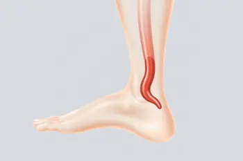
Deep vein thrombosis (DVT) is defined as the formation of a blood clot within a vein located deep inside the body, most frequently occurring in the large veins of the lower leg and thigh 1. While DVT can manifest in the arms or pelvis, the lower extremities remain the most common site of development 2. Clinical observations indicate that symptoms are often localized to one side of the body, which serves as a significant indicator for healthcare providers during initial assessments 3. Patients frequently report localized swelling, which may be accompanied by a sensation of heaviness or persistent aching in the affected limb 4.
Physical examination often reveals visible changes in the skin overlying the affected area, including redness or localized discoloration 5. Furthermore, the skin may feel noticeably warm to the touch, and individuals may experience tenderness or localized pain, which is sometimes described as cramping that exacerbates when standing or walking 6. It is a clinical reality that not all DVT cases present with overt physical symptoms; some clots are classified as silent, meaning they do not produce recognizable physical signs for the patient 7.
Understanding the Progression to Pulmonary Embolism
The primary concern regarding DVT involves the potential for the clot, or a portion thereof, to detach from its original site and travel through the bloodstream 8. This process is termed thromboembolism, and if the mobile clot becomes lodged in the pulmonary arteries, it results in a pulmonary embolism (PE) 9. PE is a medical emergency that can lead to significant respiratory distress, strain on the heart, and in severe instances, fatal outcomes 10. The movement of clots is the defining factor that transitions a localized DVT into a systemic, life-threatening condition 11.
Emergency warning signs associated with pulmonary embolism include the sudden onset of shortness of breath and chest pain that may worsen during deep inhalation or coughing 12. Additional clinical indicators of a PE include an elevated heart rate, unexplained coughing-at times involving blood-lightheadedness, or fainting 13. Because these symptoms signify a blockage of blood flow to the lungs, immediate professional medical intervention is required upon their appearance 14. Clinical protocols emphasize that DVT and PE are part of a continuum known as venous thromboembolism (VTE) 15.
Risk Factors and Demographic Predispositions
While DVT can affect individuals across all age groups, statistics indicate that the risk increases significantly for adults over the age of 60 16. The likelihood of clot formation is heightened by factors that impede normal blood flow, such as prolonged sedentary periods, including bed rest or extended travel involving long durations of sitting without movement 17. Other recognized risk factors include recent surgical procedures, particularly those involving the hips, pelvis, or legs, as well as the presence of physical trauma such as fractures in those regions 18.
Additional physiological and medical conditions also contribute to an increased probability of DVT, including obesity, pregnancy, and the six-month postpartum period 19. Furthermore, patients with chronic illnesses, including certain types of cancer, or those with indwelling catheters-tubes placed within blood vessels for medical access-face higher clinical risks 20. Hematological conditions like polycythemia vera, which causes the bone marrow to produce excessive blood cells, are also linked to elevated clot formation risks 21.
Diagnostic Processes and Medical Evaluation
Because the physical symptoms of DVT can mimic other musculoskeletal or inflammatory conditions, healthcare providers cannot confirm a diagnosis based on visual inspection alone 22. Diagnostic verification typically requires objective imaging or laboratory testing 23. The most common diagnostic tool is a Doppler ultrasound, which uses sound waves to visualize blood flow within the veins and detect the presence of obstructive clots 24.
Laboratory testing, such as the D-dimer blood test, may also be utilized during the diagnostic process. A negative D-dimer result is often used to effectively rule out the presence of a clot in low-probability cases, while a positive result is considered non-specific, indicating the need for further, more definitive imaging to confirm the presence of a thrombus 25. In specialized scenarios, clinicians may utilize venography, an invasive procedure involving the injection of contrast dye into the veins followed by X-ray imaging to map blood flow and identify blockages 26.
Management and Therapeutic Strategies
The standard treatment for confirmed DVT involves the administration of anticoagulants, commonly referred to as blood thinners 27. The primary objective of these medications is to prevent the existing clot from increasing in size and to minimize the risk of the clot breaking free and traveling to the lungs 28. Common anticoagulation agents include heparin, often administered intravenously in clinical settings, and oral medications like warfarin, though clinical monitoring is required to manage their effects 29.
Long-term health consequences of DVT can include post-phlebitis syndrome, a condition where the vein is permanently damaged by the clot 30. This syndrome may manifest as persistent leg pain, swelling, chronic skin discoloration, and in some instances, the development of skin ulcers that are difficult to heal 31. Consistent with clinical guidelines, treatment is focused on both acute stabilization and the prevention of chronic venous insufficiency, which can occur when the body struggles to return blood to the heart effectively after a clotting event 32.
Prevention and Maintaining Vascular Health
Preventative strategies are essential for individuals identified as having high-risk factors for venous thromboembolism. Recommendations often include early mobilization following surgery or illness, maintaining adequate hydration, and implementing risk-based prevention strategies during long-distance travel 33. For hospitalized patients, particularly those in intensive care units, clinicians often employ mechanical or pharmacological prophylaxis to mitigate the incidence of DVT 34.
Understanding that up to one-third of hospitalized patients may show evidence of DVT upon screening highlights the importance of vigilance in clinical environments 35. Because medical history, such as a family predisposition to clotting disorders, plays a critical role in individual risk profiles, individuals are encouraged to review their health history with medical professionals to develop personalized prevention plans 36. Ongoing research continues to focus on refining these preventative measures to reduce the morbidity associated with venous blood clots 37.
Sources
- Penn Medicine: Deep Vein Thrombosis Symptoms and Causes
- Merck Manual Consumer Version: Deep Vein Thrombosis (DVT)
- UC Davis Health: Symptoms, Dangers, and When to Get Help
- Cleveland Clinic: Don’t Ignore These 8 Blood Clot Warning Signs
- Harvard Health: Leg Clots and DVT Health Hazards
- Family Doctor: Deep Vein Thrombosis
- Nebraska Medicine: How Do You Know If You Have a Blood Clot?
- MSD Manual Professional Edition: Deep Venous Thrombosis (DVT)
- Acibadem Hospitals Group: Deep Vein Thrombosis Signs and Treatment
- Swedish Health: Understanding Deep Vein Thrombosis
- Centers for Disease Control and Prevention: DVT Symptoms
- National Heart, Lung, and Blood Institute: VTE Symptoms
- MedlinePlus: Deep Vein Thrombosis
- Mayo Clinic: DVT Symptoms and Causes
- National Health Service: DVT Symptoms and Advice
- Johns Hopkins Medicine: Deep Vein Thrombosis Information
- American Heart Association: Venous Thromboembolism Details
- Mount Sinai: DVT Warning Signs
Authored by MistyMountainView team